Myofascial Release (MFR) and Instrument-Assisted Soft Tissue Mobilization (IASTM) are manual therapy approaches that target the fascial system, the continuous three-dimensional connective tissue network that surrounds, supports, and connects every muscle, tendon, ligament, nerve, blood vessel, and organ in the body. When fascia becomes restricted, adhered, fibrotic, or disorganized due to injury, surgery, chronic overuse, immobilization, or inflammation, it can limit mobility, alter movement mechanics, compress neurovascular structures, and contribute to persistent pain.
Myofascial Release uses sustained manual pressure and slow, controlled stretching applied directly to fascial restrictions to restore tissue extensibility, fluid dynamics, and mechanotransduction signaling within the connective tissue matrix. IASTM uses specially designed rigid instruments, typically stainless steel, to detect and treat soft tissue dysfunction with greater precision, deeper penetration, and more targeted mechanical input than manual techniques alone.
At Shiva Physical Therapy, myofascial release and IASTM are applied as clinical interventions within a comprehensive regenerative framework. They are not used as generic soft tissue massage or routine warm-up techniques. They are applied with specific clinical reasoning to address identified fascial restrictions, scar tissue adhesions, and extracellular matrix dysfunction that may be limiting tissue healing, neurovascular function, and movement recovery.
Both myofascial release and IASTM work through the principle of mechanotransduction, the process by which mechanical forces applied to tissue are converted into cellular and biochemical responses. When controlled pressure, shear force, or sustained stretch is applied to restricted fascial tissue, fibroblasts within the extracellular matrix detect the mechanical stimulus and respond by initiating a cascade of biological events that support tissue repair, remodeling, and restoration.

Break down fascial adhesions, scar tissue, and fibrotic restrictions that limit tissue mobility
Stimulate fibroblast proliferation and activation, increasing the production of new collagen and extracellular matrix proteins
Promote collagen synthesis, realignment, and maturation, improving the structural organization and tensile strength of healing tissue
Increase local microcirculation, blood flow, and nutrient delivery to restricted or hypoxic tissue
Restore extracellular matrix mobility and fluid dynamics within fascial layers
Stimulate mechanoreceptors in the skin and fascia, influencing pain perception and neuromuscular activation
Reduce muscle guarding, protective tension, and compensatory movement patterns
Improve proprioceptive feedback and sensory awareness in treated regions
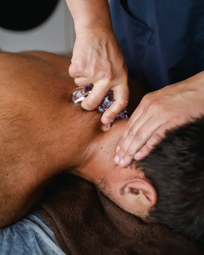
IASTM provides an additional mechanical advantage. The rigid instruments amplify the clinician's ability to detect subtle tissue changes, including areas of increased density, fibrosis, or fascial restriction, through vibration and texture feedback transmitted through the tool. This allows for more precise identification and treatment of dysfunctional tissue compared to manual palpation alone.
When applied with clinical precision and integrated within a structured rehabilitation framework, myofascial release and IASTM serve as powerful connective tissue restoration tools that may help prepare the fascial environment for more effective loading, strengthening, and functional recovery.
Many manual therapy approaches focus primarily on muscle tension, joint mobility, or spinal alignment. Myofascial release and IASTM work at the fascial and extracellular matrix level, targeting the connective tissue system that influences how muscles, nerves, and blood vessels function within their structural environment.
Myofascial Release and IASTM are different because they:
Target the fascial system directly, not just the muscles or joints it surrounds
Influence tissue biology at the cellular level through mechanotransduction and fibroblast activation
Address scar tissue, adhesions, and fibrotic restrictions that other manual techniques may not effectively reach
Restore extracellular matrix organization and fluid dynamics within connective tissue layers
Provide both diagnostic and therapeutic value, as instruments can detect tissue abnormalities while simultaneously treating them
Support tissue remodeling and collagen reorganization, not just temporary symptom relief
Myofascial release and IASTM offer a way to influence the connective tissue environment that determines how tissue heals, how forces are transmitted through the body, and how freely structures move within their fascial compartments. By restoring fascial health, these techniques may help create conditions where other interventions, including exercise, loading, and regenerative technologies, can be more effective.
Research and laboratory studies suggest that myofascial release and IASTM may contribute to several biological and cellular responses:

Increased fibroblast proliferation and activation within the extracellular matrix
Enhanced collagen synthesis, fiber alignment, and structural maturation in healing tissue
Increased fibronectin production, a glycoprotein essential for cell adhesion, tissue formation, and repair
Resorption of excessive fibrosis and breakdown of disorganized scar tissue
Increased local tissue temperature and metabolic activity
Stimulation of mechanoreceptors, influencing proprioception, two-point discrimination, and pain perception
Modulation of mechanonociceptor activity, potentially reducing pain sensitivity in treated areas
Reduction of muscle guarding, myofascial trigger point activity, and protective tension
Improved fascial glide and inter-layer sliding between adjacent tissue planes
Enhanced lymphatic flow and reduction of local edema
These mechanisms may help create a more favorable connective tissue environment for healing when combined with progressive loading, therapeutic exercise, and other regenerative interventions.
Myofascial release and IASTM are used to support the body's natural tissue repair and remodeling processes by directly influencing the fascial and connective tissue structures that play a critical role in mobility, force transmission, and pain regulation. These techniques do not cure disease. They help restore the structural and biological conditions under which tissue can function, heal, and adapt more effectively.
Pain reduction, particularly in conditions involving fascial restriction or scar tissue
Improved range of motion and joint mobility
Decreased muscle tension and myofascial tightness
Improved tissue extensibility and flexibility
Enhanced recovery from injury, surgery, or immobilization
Reduction of scar tissue adhesions and post-surgical stiffness
Improved movement quality and reduced compensatory patterns
Enhanced neuromuscular activation and proprioceptive awareness
Better response to therapeutic exercise and progressive loading
Each person's response depends on factors such as the nature and chronicity of their condition, tissue quality, overall health status, and how these techniques are integrated within their broader treatment plan. Results are individual and not guaranteed.
Myofascial release and IASTM encompass a range of manual and instrument-based approaches, each designed to address specific types of fascial dysfunction. The techniques selected depend on clinical findings during evaluation, tissue irritability, treatment goals, and the phase of recovery.

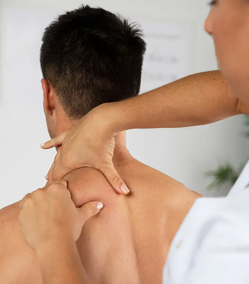
Manual myofascial release involves applying sustained, low-load pressure and slow stretching directly to fascial restrictions using the clinician's hands. Pressure is maintained until the tissue barrier is felt to release, allowing the fascia to elongate, rehydrate, and restore normal extensibility. This technique is particularly effective for broad fascial restrictions, deep tissue holding patterns, and conditions where gentle, sustained input is more appropriate than higher-force mobilization. Sustained holds typically last 90 to 120 seconds or longer, allowing time for the viscoelastic properties of fascia to respond and the tissue to reorganize.
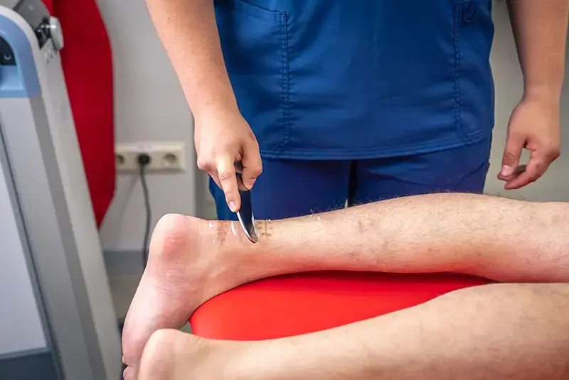
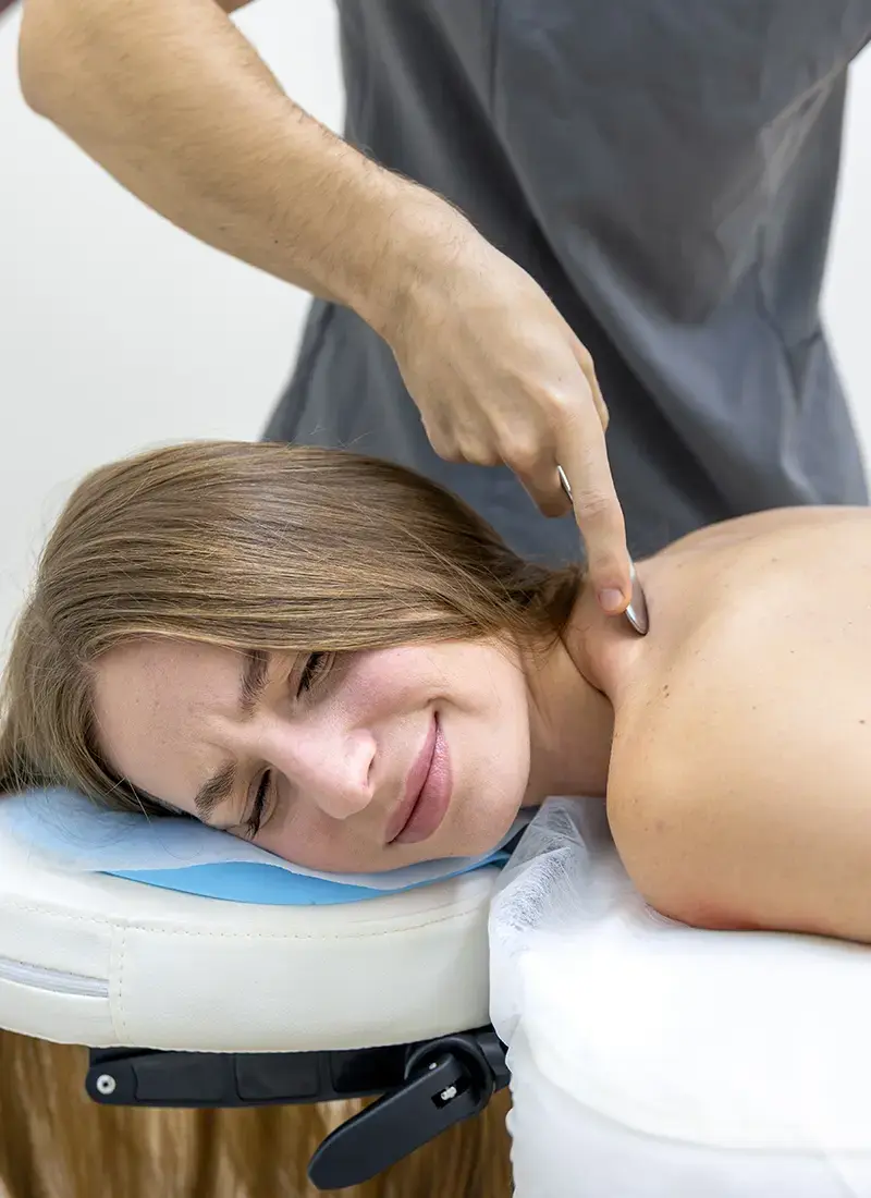
IASTM uses rigid, ergonomically designed instruments, typically made from stainless steel, to apply controlled mechanical force to soft tissue. The instruments allow the clinician to detect areas of fascial restriction, scar tissue, fibrosis, and adhesion through tactile feedback transmitted through the tool, and to treat those areas with precise, directed mechanical input. IASTM is applied at specific angles and pressures, typically for 40 to 120 seconds per treatment area, and is always followed by movement and loading to reinforce the tissue changes achieved during treatment. Common IASTM systems used clinically include various stainless steel instrument sets designed for different body regions and tissue depths.
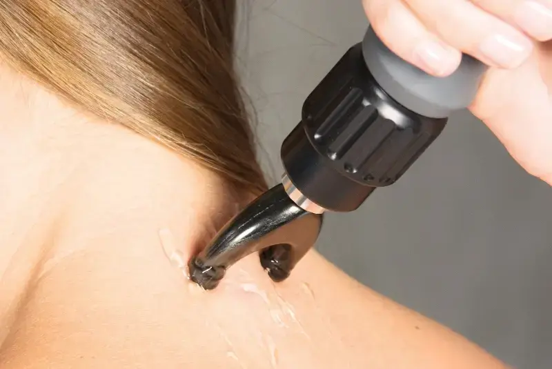
Deep fascial release targets the deeper fascial layers, including the deep investing fascia, intermuscular septa, and periosteal fascial attachments. These deeper restrictions can develop from chronic overuse, surgical scarring, or prolonged immobilization and may contribute to persistent stiffness, restricted joint motion, and neurovascular compression. Deep fascial techniques require advanced palpation skills and careful clinical reasoning to apply appropriate force without excessive tissue irritation.

Based on the work of Luigi Stecco and the Fascial Manipulation model, fascial densification techniques target specific points within the fascial system where the loose connective tissue (hyaluronic acid-rich ground substance) between fascial layers has become densified and lost its normal fluid properties. When these areas densify, fascial layers lose their ability to glide smoothly over one another, leading to altered mechanotransduction, restricted movement, and pain. Treatment involves applying deep, sustained friction to restore the viscosity and gliding properties of the interfascial matrix.
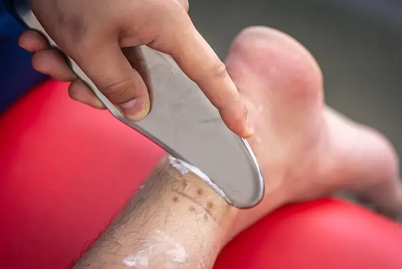
Following surgery, trauma, or significant tissue injury, scar tissue can form in ways that restrict mobility, compress neurovascular structures, and disrupt the normal organization of collagen fibers within the extracellular matrix. Scar tissue mobilization uses both manual and instrument-based techniques to break down excessive adhesions, promote collagen realignment, improve local circulation, and restore functional tissue extensibility. This is particularly important for post-surgical patients or individuals with chronic conditions involving significant fibrotic changes.
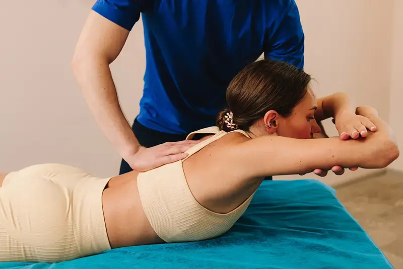
Fascia surrounds and envelops peripheral nerves and blood vessels throughout the body. When fascial restrictions develop around neurovascular structures, they can contribute to nerve compression, reduced blood flow, altered nerve conduction, and symptoms such as numbness, tingling, burning, or radiating pain. Neurovascular fascial release techniques use gentle, sustained manual pressure along the pathways of affected nerves and vessels to restore fascial mobility and reduce mechanical compression on these structures.
Not every patient requires all techniques. The choice depends on individual evaluation findings, clinical presentation, tissue reactivity, treatment goals, and the phase of recovery within the overall treatment plan.
Myofascial release and IASTM are commonly considered when clinical evaluation suggests that fascial restriction, scar tissue adhesion, extracellular matrix dysfunction, or connective tissue fibrosis may be contributing to the patient's pain, movement limitation, or impaired recovery.


Chronic neck and back pain
Shoulder impingement and frozen shoulder (adhesive capsulitis)
Plantar fasciitis
Lateral and medial epicondylitis (tennis elbow, golfer's elbow)
IT band syndrome
Patellar tendinopathy
Achilles tendinopathy
Post-surgical scar tissue and adhesions
Rotator cuff injuries and post-repair stiffness
Myofascial pain syndrome and trigger points
Carpal tunnel syndrome and nerve entrapment conditions
Chronic muscle strains and recurrent soft tissue injuries
Post-fracture stiffness and joint contracture
Thoracolumbar fascia restrictions
Sports injuries involving fascial and connective tissue damage
Reduced range of motion due to fibrotic changes or immobilization
Not every condition or patient requires myofascial release or IASTM. A thorough clinical assessment helps determine whether these techniques are appropriate as part of an individual's treatment plan.
Myofascial release and IASTM are not applied routinely or universally to every patient. Within The S.H.I.V.A. Method, their use is determined by clinical evaluation, individual presentation, and the specific fascial and connective tissue needs identified during assessment.
Our approach focuses on when to apply these techniques, which approach to use, and when other interventions are more appropriate. This includes decisions such as:
Because fascial tissue responds to sustained and repeated mechanical input over time, treatment timing and sequencing matter. Applying myofascial release or IASTM at the right phase of healing can accelerate tissue remodeling and prepare the connective tissue environment for loading. Applied at the wrong time or with excessive force, it can increase irritation and delay recovery.
Within The S.H.I.V.A. Method, myofascial release and IASTM serve as components of a comprehensive strategy that also addresses:
When used appropriately, these techniques can be valuable contributors to regenerative physical therapy (RPT) by helping restore the fascial and connective tissue environment that influences how tissue heals, how forces are transmitted, and how the body responds to rehabilitation.
Myofascial release and IASTM sessions are targeted, focused, and designed to support connective tissue restoration and movement recovery. The experience differs depending on which technique is applied.
Myofascial release and IASTM techniques are typically applied for 10 to 20 minutes as part of a comprehensive treatment session.
Most patients receive these techniques 1 to 2 times per week as part of a structured rehabilitation plan. Frequency is adjusted based on individual tissue response and the phase of recovery.
A typical course ranges from 1 to 4sessions, depending on the nature and chronicity of the fascial dysfunction. The treatment cycle is adjusted based on tissue response and evolving clinical goals.
Sensation (IASTM): You may feel a scraping, rubbing, or friction sensation as the instrument moves across the skin. Treatment should never produce too sharp pain or excessive bruising.
Minimal preparation is required. Clothing should allow access to the treatment area. A topical emollient may be applied to facilitate instrument glide during IASTM.
There is no required downtime. Most patients resume normal activities immediately. Mild soreness in treated areas is common and typically resolves within 24 to 48 hours..
Some patients notice improvements in mobility, tissue flexibility, or pain within the first few sessions, with continued progress as treatment is integrated with progressive loading and other components of their rehabilitation plan.
Myofascial release and IASTM are considered when clinical evaluation suggests that fascial restriction, scar tissue adhesion, or connective tissue dysfunction may be contributing to the patient's pain, movement limitation, or impaired recovery.
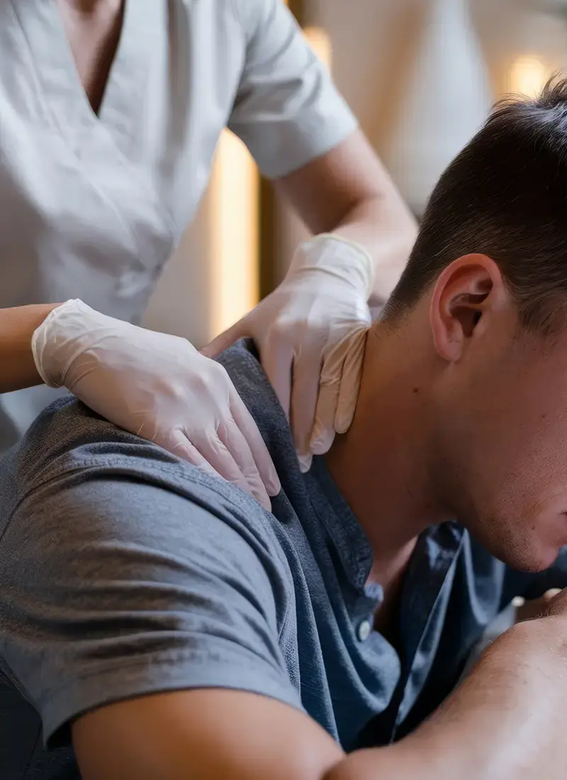
These techniques may be appropriate for individuals who:
Have chronic soft tissue tightness, restricted mobility, or persistent stiffness despite stretching and exercise
Present with post-surgical scar tissue or adhesions limiting functional recovery
Experience recurrent soft tissue injuries suggesting underlying fascial restriction or tissue disorganization
Have tendinopathy, fasciitis, or connective tissue conditions with fibrotic changes
Show signs of fascial densification, restricted fascial glide, or altered tissue texture on clinical examination
Need fascial preparation before progressive loading and strengthening can be effective
Have chronic pain associated with myofascial trigger points, muscular holding patterns, or regional soft tissue dysfunction
Are athletes or active individuals requiring tissue restoration and optimized fascial mobility for performance
Clinical screening determines suitability. The decision to use myofascial release or IASTM is always based on individual findings, overall clinical reasoning, and the patient's specific treatment goals.
Myofascial release is a hands-on manual technique that uses sustained pressure and slow stretching to release fascial restrictions. IASTM uses specially designed rigid instruments to apply controlled mechanical force to soft tissue, providing deeper penetration, greater precision, and enhanced ability to detect tissue abnormalities through tactile feedback. Both approaches target the fascial system and connective tissue, but they offer different mechanical advantages depending on the clinical situation. Your clinician will determine which approach, or combination, is most appropriate for your condition.
Instrument-assisted soft tissue mobilization: a systematic review and effect-size analysis.
Read Full StudyEffectiveness of myofascial release for adults with chronic neck pain: a meta-analysis.
Read Full StudyMyofascial release for the treatment of tension-type, cervicogenic headache or migraine: a systematic review and meta-analysis.
Read Full StudyEffectiveness of myofascial release: systematic review of randomized controlled trials.
Read Full Study
Effects of myofascial release techniques on joint range of motion of athletes: a systematic review and meta-analysis of randomized controlled trials.
Read Full StudyMyofascial release strategies and technique recommendations for athletic performance: a systematic review.
Read Full StudyTherapeutic effectiveness of instrument-assisted soft tissue mobilization for soft tissue injury: mechanisms and practical application.
Read Full StudyInstrument-assisted soft-tissue mobilization: a commentary on clinical practice guidelines for rehabilitation professionals.
Read Full StudyInstrument-assisted cross-fiber massage increases tissue perfusion and alters microvascular morphology in the vicinity of healing knee ligaments.
Read Full Study